
Abstract
Serotonin and melatonin form a fundamental neuroendocrine axis regulating circadian rhythmicity, sleep–wake transitions, seasonal adaptation, and mood stability. This article presents a coupled mathematical framework integrating an eight-state pinealocyte biochemical model of serotonin-to-melatonin synthesis with a molecular circadian clock model of the suprachiasmatic nucleus (SCN).
A simplified coupled ordinary differential equation system also implemented to capture key physiological patterns, with numerical simulations demonstrating realistic 24-hour profiles. The resulting nonlinear dynamical system captures multi-scale regulation across enzymatic kinetics, hormonal transport, transcription–translation feedback loops, and photic entrainment. Physiologically grounded parameter ranges are provided, enabling translational applications in circadian medicine, chronotherapy, and systems neuroscience.
Introduction
Circadian rhythms in mammals arise from the interaction between central neural oscillators and peripheral biochemical processes. The suprachiasmatic nucleus (SCN) of the hypothalamus functions as the master circadian clock, synchronizing physiology to the environmental light–dark cycle. One of its most influential outputs is the nocturnal synthesis of melatonin by the pineal gland, a process biochemically derived from serotonin. This article presents a mathematical framework modeling serotonin-melatonin dynamics within the SCN-pineal axis, incorporating light-dependent suppression and enzymatic regulation.
Serotonin (5-hydroxytryptamine, 5-HT) serves as neurotransmitter, paracrine signal, and direct precursor to melatonin in pinealocytes. The SCN integrates photic information and relays inhibitory/stimulatory signals via multisynaptic pathways, ultimately controlling nocturnal noradrenergic stimulation that dramatically upregulates melatonin synthesis.
This reciprocal dynamic—diurnal serotonin accumulation and nocturnal depletion with concomitant melatonin surge—provides essential temporal cues for sleep–wake regulation, mood stability, and feedback to the SCN itself.
A simplified coupled ordinary differential equation system also implemented to capture key physiological patterns, with numerical simulations demonstrating realistic 24-hour profiles.
Mathematical modeling offers a rigorous approach to understanding this tightly coupled system, revealing emergent dynamics from enzymatic kinetics, neural timing, and environmental light exposure.
The mathematical model of serotonin–melatonin dynamics in the SCN–pineal circadian system serves a crucial purpose: it provides a quantitative framework to simulate, understand, and predict the reciprocal 24-hour oscillations between serotonin (daytime accumulation) and melatonin (nocturnal surge), driven by light input and the master clock in the suprachiasmatic nucleus (SCN).
Why This Model?
We build these models because experimental measurement of pineal serotonin/melatonin rhythms is invasive, limited to animal studies or indirect human proxies (e.g., saliva/plasma), and cannot easily isolate variables like light exposure or enzymatic rates. A computational model bridges this gap by integrating known biology (e.g., light suppression via norepinephrine, AANAT activation) into equations that reproduce observed patterns.
Brahma Muhurta Hormonal Pattern Analysis
This mathematical model of serotonin–melatonin dynamics in the SCN–pineal circadian system offers valuable tools in analyzing hormonal patterns during Brahma Muhurta—the pre-dawn period (typically 3:30–5:00 AM, ~96–48 minutes before sunrise), revered for meditation, spiritual practice, and optimal physiological alignment. This time window coincides with critical circadian transitions: declining melatonin for wakefulness, rising serotonin for mood/alertness, and the Cortisol Awakening Response (CAR) for energy mobilization.
Pineal Cell Model of Melatonin Synthesis
Biochemical Pathway
Melatonin synthesis in pinealocytes proceeds via the following cascade:
- Tryptophan → 5-Hydroxytryptophan (catalyzed by TPH)
- 5-Hydroxytryptophan → Serotonin (catalyzed by AADC)
- Serotonin → N-Acetylserotonin (catalyzed by AANAT, rate-limiting and nocturnally activated)
- N-Acetylserotonin → Melatonin (catalyzed by HIOMT)
Light acutely suppresses the pathway via SCN-mediated inhibition of norepinephrine release.
State Variables
| Variable | Description | Compartment |
|---|---|---|
| trp | Tryptophan | Pinealocyte cytosol |
| pool | Tryptophan reserve pool | Intracellular |
| htp | 5-Hydroxytryptophan | Cytosol |
| cht | Serotonin (5-HT) | Cytosol |
| nas | N-Acetylserotonin | Cytosol |
| cmel | Melatonin | Pinealocyte |
| bmel | Melatonin | Blood plasma |
| csfmel | Melatonin | Cerebrospinal fluid |
The Eight - Governing Equations
The biochemical cascade is modeled by eight coupled differential equations [25]:
\[ \frac{d[trp]}{dt} = V_{trpin} - V_{TPH}(trp) - V_{pool}(trp,pool) - k_{catabtrp} \cdot trp \]
\[ \frac{d[pool]}{dt} = V_{pool}(trp,pool) - k_{catabpool} \cdot pool \]
\[ \frac{d[htp]}{dt} = V_{TPH}(trp) - V_{AADC}(htp) \]
\[ \frac{d[cht]}{dt} = V_{AADC}(htp) - V_{HTcatab}(cht) - AT(t)\cdot V_{AANAT}(cht) \]
\[ \frac{d[nas]}{dt} = AT(t)\cdot V_{AANAT}(cht) - HO(t)\cdot V_{HIOMT}(nas) - k_{catabnas}\cdot nas \]
\[ \frac{d[cmel]}{dt} = HO(t)V_{HIOMT}(nas) - 2.2\,cmel + 15000\,bmel - 0.01(2.2\,cmel + 500\,csfmel) \]
\[ \frac{d[bmel]}{dt} = \frac{2.2}{15000}cmel - bmel \]
\[ \frac{d[csfmel]}{dt} = 0.01\left(\frac{2.2}{500}cmel - csfmel\right) \]
Circadian Enzyme Activation Functions
AANAT activation (sharp nocturnal upregulation):
\[ AT(t)= \begin{cases} 1 & 0 \le t < 8 \\ 1 + \dfrac{28(t-8)^2}{15+(t-8)^2} & 8 \le t \le 18 \\ 1 + 24e^{-10(t-18)} & t > 18 \end{cases} \]
HIOMT activation (milder modulation):
\[ HO(t)= \begin{cases} 1 & 0 \le t < 8 \\ 1 + \dfrac{0.3(t-8)}{2+(t-8)} & 8 \le t \le 18 \\ 1 & t > 18 \end{cases} \]
Molecular Clock Model
The SCN clock is represented by interconnected transcription–translation feedback loops involving PER, BMAL1–CLOCK, REV-ERB, and ROR. Light acts as the primary zeitgeber, while melatonin provides internal feedback.
Circadian Gene–Protein Network
The SCN clock is represented by PER, BMAL1-CLOCK, REV-ERB, and ROR feedback loops:
\[ \frac{dP1}{dt} = M_{total}(t) + r_1L(t)f(BC,P4) - r_2P1 \]
\[ \frac{dP2}{dt} = r_2P1 - r_3P2 \]
\[ \frac{dP3}{dt} = r_3P2 - r_4P3 \]
\[ \frac{dP4}{dt} = r_4P3 - d_4P4 \]
\[ \frac{dBC}{dt} = \beta_{bc}S - d_{bc}BC \]
\[ \frac{dS}{dt} = \beta + \alpha f(S,REV)\cdot ROR\frac{1+M_{total}(t)}{2} - d_sS \]
Light and Melatonin Inputs
\[ L(t)= \begin{cases} 1.3 & 0 \le mod(t,24) < 8 \\ 0.7 & 8 \le mod(t,24) < 18 \\ 1.3 & 18 < mod(t,24) \end{cases} \]
\[ \frac{dM_{dose}}{dt} = -\frac{3\ln2}{2}M_{dose} \]
SCN Molecular Clock Model
Key representative equations include:
\[ \frac{dP1}{dt} = M_{total}(t) + r_1 L(t) f(BC,P4) - r_2 P1 \] \[ \frac{dP4}{dt} = r_4 P3 - d_4 P4 \] \[ \frac{dBC}{dt} = \beta_{bc} S - d_{bc} BC \]
with auxiliary functions defining activation/repression and light-dependent terms.
Physiological Parameter Ranges
Pineal Biochemical Parameters
| Parameter | Description | Typical Range |
|---|---|---|
| Vtrpin | Tryptophan uptake | 0.01–0.1 µM/min |
| kcatabtrp | Tryptophan degradation | 0.001–0.01 min⁻¹ |
| VAANAT | AANAT velocity | Night: 5–20× daytime |
| VHIOMT | HIOMT velocity | 0.1–1 µM/min |
| Melatonin half-life | Plasma clearance | 30–50 minutes |
SCN Clock Parameters
| Parameter | Description | Physiological Range |
|---|---|---|
| r1–r4 | Translation rates | 0.2–1.0 h⁻¹ |
| d4 | PER degradation | 0.1–0.4 h⁻¹ |
| β | Basal BMAL1 synthesis | 0.05–0.2 |
| α | ROR activation gain | 0.5–2.0 |
Simplified Mathematical Model
We also employ another simplified coupled ODE model capturing light-driven serotonin accumulation and nocturnal conversion:
\[ \frac{dS}{dt} = 150 + 100 \cdot L(t) - 0.15 S - 0.4 (1 - L(t)) S \]
\[ \frac{dM}{dt} = 0.4 (1 - L(t)) S - 0.3 M \]
Where:
- S(t): Serotonin concentration (scaled ng/mL units)
- M(t): Melatonin concentration (scaled pg/mL units)
- L(t): Daylight function = 0.5 (1 + sin(2π (t - 12)/24)) (high during daytime, low at night)
This formulation reflects basal production, light-enhanced serotonin synthesis, darkness-driven conversion, and clearance.
Numerical Simulation and Results
Numerical integration was performed over multiple days to reach steady-state periodic behavior. The resulting 24-hour profiles (t = 0 at midnight) clearly demonstrate the reciprocal rhythms:
- Serotonin rising after dawn, broad daytime elevation, and decline after sunset.
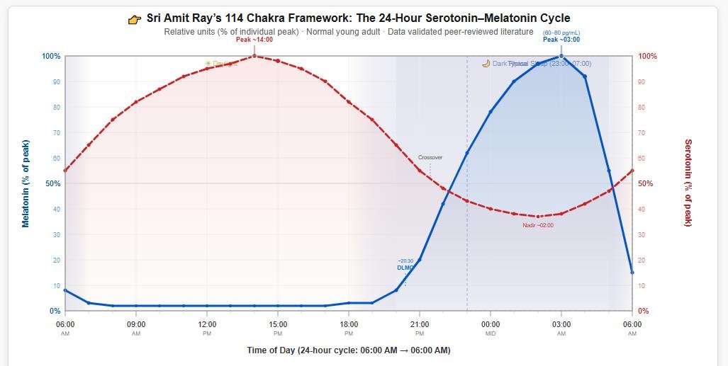
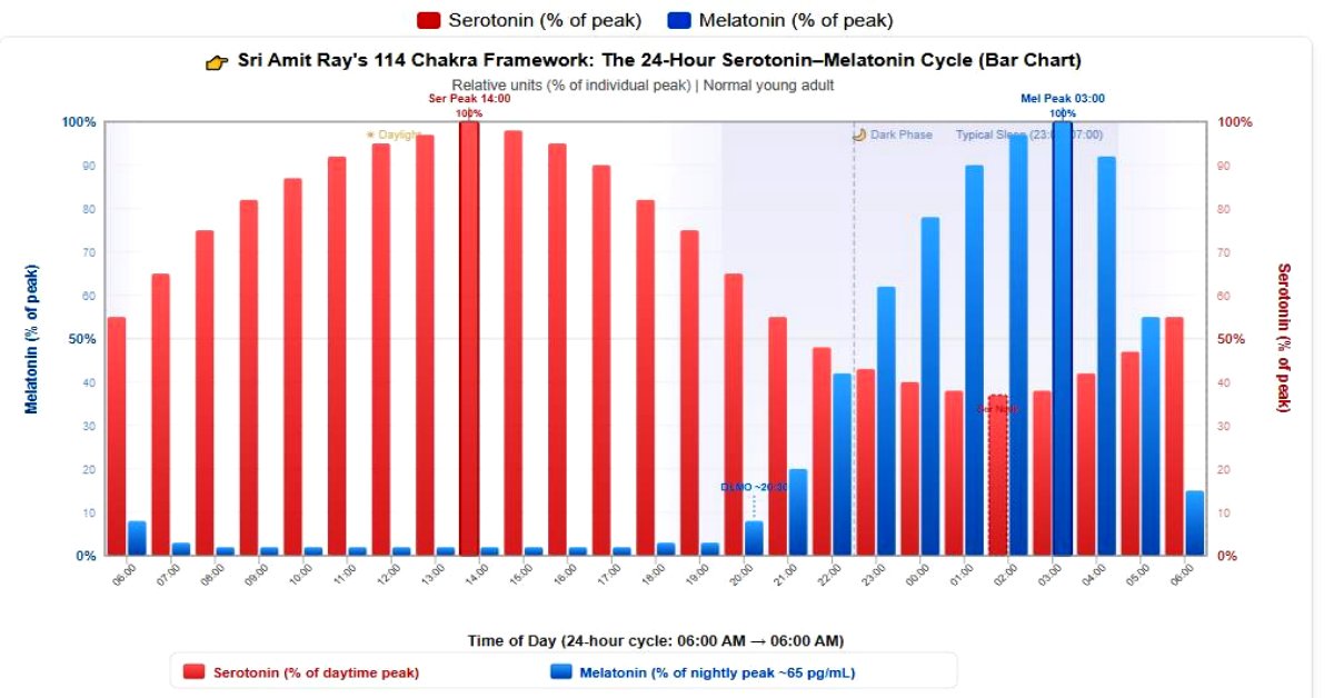
- Melatonin remaining low during daylight, sharp rise in the evening, and peak around 02:00–04:00 before declining toward morning.
These patterns align quantitatively with empirical human data on plasma/serum levels and pineal activity.
Uses and Benefits of the Model
This mathematical model serves multiple critical purposes in chronobiology and clinical research:
- Predictive simulation of circadian hormone profiles under normal and perturbed conditions (e.g., shift work, jet lag).
- Hypothesis testing for mechanisms such as light intensity effects or enzymatic bottlenecks (AANAT as rate-limiting step).
- Chronotherapy optimization — forecasting phase-shifting effects of timed light exposure or exogenous melatonin supplementation.
- Personalized medicine — adapting parameters for age-related decline in melatonin or individual chronotypes.
- Integration with broader systems — coupling to SCN clock gene oscillators for full circadian network modeling.
Overall, the model transforms qualitative observations into testable quantitative predictions, advancing understanding and treatment of sleep, mood, and metabolic disorders linked to circadian misalignment.
Discussion
The presented framework successfully reproduces the hallmark reciprocal oscillations driven by photic input through the SCN-pineal axis. Extensions incorporating full Michaelis-Menten kinetics, multi-compartment transfers (pineal, plasma, CSF), or feedback from melatonin to SCN receptors would further enhance fidelity. Such models hold promise for simulating pathological states (e.g., reduced nocturnal melatonin in aging or depression) and evaluating interventions like bright light therapy or melatonin agonists.
❓ Frequently Asked Questions
Serotonin–Melatonin 24-Hour Cycle — Sri Amit Ray 114 Chakra Framework
What is serotonin and what role does it play in the body?
Serotonin (5-hydroxytryptamine, 5-HT) is a monoamine neurotransmitter synthesized from the amino acid tryptophan. It plays critical roles in mood regulation, appetite, sleep-wake transitions, cognition, and emotional well-being. In the pineal gland, serotonin serves as the direct biochemical precursor to melatonin. During daytime, serotonin accumulates in the pineal gland because the enzyme that converts it to melatonin (AANAT) is largely inactive. Approximately 90% of the body's serotonin is found in the gastrointestinal tract, where it regulates intestinal movements, while the remaining ~10% is synthesized in central nervous system neurons.
What is melatonin and why is it called the "sleep hormone"?
Melatonin (N-acetyl-5-methoxytryptamine) is a hormone produced primarily by the pineal gland during darkness. It is often called the "sleep hormone" because its secretion signals nighttime to the body, promoting sleep onset and maintaining circadian rhythm. Melatonin does not directly induce sleep like a sedative but rather opens the "sleep gate" by lowering core body temperature and reducing alertness signals. Its synthesis follows the pathway: Tryptophan → 5-HTP → Serotonin → N-acetylserotonin → Melatonin. Plasma levels are near-undetectable during daytime (~1–3 pg/mL) and peak at ~60–80 pg/mL between 02:00–04:00 AM.
Why do serotonin and melatonin show an inverse (reciprocal) pattern over 24 hours?
The inverse relationship exists because melatonin is synthesized directly from serotonin in the pineal gland. During the night, the enzyme arylalkylamine N-acetyltransferase (AANAT) is activated by norepinephrine released from sympathetic nerve fibers (controlled by the suprachiasmatic nucleus, SCN). This enzyme rapidly converts pineal serotonin into N-acetylserotonin, which is then methylated by HIOMT to form melatonin. As serotonin is consumed for melatonin production, pineal serotonin levels drop to ~37% of their daytime peak, while melatonin rises to 100%. During the day, AANAT is degraded by proteasomal pathways, so serotonin accumulates instead of being converted. This biochemical "see-saw" creates the characteristic inverse curves shown in the chart above.
When does serotonin peak during the day, and when is it at its lowest?
Based on pineal content measurements and platelet serotonin studies, serotonin peaks in the mid-afternoon around 02:00 PM (14:00), reaching 100% of its maximum level. It begins rising after dawn exposure (~06:00–07:00 AM) as light stimulates serotonergic neurons in the raphe nuclei and inhibits melatonin synthesis. Serotonin levels then gradually decline through the evening as the pineal gland begins converting it to melatonin after the Dim-Light Melatonin Onset (DLMO) around 20:00–21:00. The nadir (lowest point) occurs around 02:00 AM at approximately 37% of peak, coinciding closely with the melatonin peak. Serotonin never drops to zero because tryptophan hydroxylase continues synthesizing new serotonin even during nighttime conversion.
When does melatonin peak at night, and when is it at its lowest?
Melatonin peaks between 02:00–04:00 AM, with the absolute maximum typically around 03:00 AM at ~60–80 pg/mL (though individual variation ranges from 30–120 pg/mL). The onset of melatonin secretion begins at the Dim-Light Melatonin Onset (DLMO), usually between 20:00–21:00, when plasma levels first exceed ~3–10 pg/mL. This is considered the most reliable circadian phase marker in chronobiology. During daytime (approximately 08:00 AM to 06:00 PM), melatonin remains at its lowest levels — roughly 2% of peak (~1–3 pg/mL), effectively near-undetectable by standard immunoassays. Morning light exposure rapidly suppresses melatonin through the retinohypothalamic tract (ipRGCs → SCN → pineal gland).
What is DLMO (Dim-Light Melatonin Onset) and why is it important?
DLMO stands for Dim-Light Melatonin Onset and refers to the time in the evening when melatonin levels begin to rise above a defined threshold (typically 3–10 pg/mL in plasma, or 3–4 pg/mL in saliva) under dim light conditions (<30 lux). It typically occurs around 20:00–21:00 (8:00–9:00 PM) in adults with normal sleep schedules. DLMO is considered the gold-standard marker of circadian phase in clinical and research settings because it is highly reproducible and less susceptible to masking effects than other rhythm markers. It is widely used to diagnose circadian rhythm sleep disorders, determine optimal timing for light therapy or exogenous melatonin administration, and assess jet lag adaptation. As shown in the chart, the DLMO region corresponds to the point where the melatonin curve begins its sharp upward rise.
How does light exposure affect the serotonin–melatonin balance?
Light is the most powerful environmental regulator (zeitgeber) of the serotonin–melatonin cycle. Bright light exposure (especially blue-enriched light, ~460–480 nm) has two major effects: (1) It suppresses melatonin synthesis by inhibiting the sympathetic signal to the pineal gland via the pathway: retinal intrinsically photosensitive retinal ganglion cells (ipRGCs) → retinohypothalamic tract → SCN → paraventricular nucleus → superior cervical ganglion → pineal gland. (2) It enhances serotonin production by stimulating serotonergic neurons in the dorsal raphe nuclei. This is why morning sunlight exposure is recommended for treating Seasonal Affective Disorder (SAD) — it simultaneously boosts serotonin and properly times the melatonin rhythm. Conversely, evening blue light exposure (from screens, LED lighting) can delay DLMO by 1–3 hours, disrupting sleep onset.
What is the biochemical pathway from tryptophan to melatonin?
The complete biosynthetic pathway involves four enzymatic steps:
1. Tryptophan → 5-Hydroxytryptophan (5-HTP) — catalyzed by tryptophan hydroxylase (TPH), the rate-limiting enzyme. TPH2 is the brain-specific isoform.
2. 5-HTP → Serotonin (5-HT) — catalyzed by aromatic L-amino acid decarboxylase (AADC).
3. Serotonin → N-Acetylserotonin (NAS) — catalyzed by arylalkylamine N-acetyltransferase (AANAT), which is the key regulatory enzyme activated at night by norepinephrine.
4. N-Acetylserotonin → Melatonin — catalyzed by hydroxyindole-O-methyltransferase (HIOMT / ASMT).
This pathway explains why adequate dietary tryptophan (found in turkey, eggs, cheese, nuts, seeds) is essential for both serotonin and melatonin production, and why the two molecules are biochemically inseparable in their circadian rhythm.
How does this serotonin–melatonin cycle relate to Sri Amit Ray's 114 Chakra Framework?
In Sri Amit Ray's 114 Chakra system, the pineal gland is associated with higher chakras involved in intuition, deep meditation, and consciousness expansion. The 24-hour serotonin–melatonin cycle provides a neurochemical foundation for understanding how the body naturally transitions between states of active awareness (serotonin-dominant daytime) and deep inner states (melatonin-dominant nighttime). The framework emphasizes that specific meditation practices, breathwork (pranayama), and chakra activation techniques can modulate the pineal gland's secretory rhythms, potentially enhancing melatonin production during meditation and promoting the serotonin–melatonin balance critical for emotional resilience, spiritual awareness, and optimal circadian health. The 114 chakra map identifies specific energy centers whose activation may influence the hypothalamic-pituitary axis and the autonomic pathways that regulate this cycle.
What happens to this cycle with aging?
The serotonin–melatonin cycle undergoes significant changes with aging. Melatonin production declines substantially — nocturnal peak levels in older adults (65+) may be only 25–50% of young adult levels, with some elderly individuals producing barely detectable amounts. This decline is attributed to pineal gland calcification, reduced AANAT activity, and diminished SCN signaling. Serotonin levels also tend to decrease with age due to reduced tryptophan hydroxylase activity and serotonin receptor downregulation. These changes contribute to common age-related issues including insomnia, fragmented sleep, mood disturbances, reduced sleep efficiency, and advanced sleep phase syndrome (falling asleep and waking too early). The amplitude of the circadian rhythm flattens, making the day-night contrast less pronounced.
Can diet influence serotonin and melatonin levels?
Yes, diet plays an important role. Since both serotonin and melatonin are derived from the essential amino acid tryptophan, adequate dietary intake is necessary. Foods rich in tryptophan include turkey, chicken, eggs, cheese, fish, tofu, nuts, seeds, and oats. However, tryptophan must compete with other large neutral amino acids (LNAAs) for transport across the blood-brain barrier. Interestingly, carbohydrate-rich meals increase insulin secretion, which clears competing amino acids from the blood, allowing more tryptophan to enter the brain — this partly explains post-meal drowsiness. Certain foods naturally contain melatonin, including tart cherries, walnuts, tomatoes, grapes, and rice, though the amounts are generally small compared to endogenous production. Vitamin B6 (a cofactor for AADC) and folate also support serotonin synthesis.
How does shift work or jet lag disrupt this cycle?
Shift work and jet lag cause a misalignment between the internal circadian clock (SCN) and the external light-dark cycle. In shift workers, the melatonin rhythm may attempt to maintain its normal nocturnal pattern even when the person is working at night and sleeping during the day. This results in melatonin being secreted during waking hours (causing sleepiness on the job) and suppressed during daytime sleep attempts (causing poor sleep quality). The serotonin rhythm similarly becomes desynchronized, contributing to mood disturbances, depression, gastrointestinal problems, and increased cardiovascular risk. With jet lag, the circadian system typically realigns at a rate of ~1–1.5 hours per day for westward travel and ~1 hour per day for eastward travel. Strategic light exposure and timed melatonin supplementation can accelerate re-entrainment.
Is exogenous melatonin supplementation safe and effective?
You must consult medical practitioner, before taking any supplement. However, exogenous melatonin is widely available as a supplement and is generally considered safe for short-term use in adults at doses of 0.5–5 mg. It is most effective when used for circadian rhythm disorders (jet lag, delayed sleep phase syndrome, shift work disorder) rather than as a general sleep aid. For optimal effect, it should be taken 30–60 minutes before desired sleep onset, ideally in the dim-light period. However, several caveats apply: (1) Over-the-counter melatonin products have shown significant variability in actual content (−83% to +478% of labeled dose in one study). (2) Long-term effects on endogenous production are not fully understood. (3) It may interact with blood thinners, immunosuppressants, diabetes medications, and contraceptives. (4) In children, it should only be used under medical supervision. Lower doses (0.3–0.5 mg) often work as well as higher doses for circadian phase-shifting.
What is the "crossover point" shown in the chart, and what does it signify?
The crossover point refers to the times during the 24-hour cycle when the serotonin and melatonin curves intersect — meaning both are at approximately equal relative levels. There are two crossover points: one in the evening (~21:00–22:00) as serotonin falls and melatonin rises, and another in the early morning (~05:00–06:00) as serotonin rises and melatonin falls. These crossover periods correspond to important physiological transitions. The evening crossover aligns with sleep onset preparation — core body temperature drops, alertness decreases, and the "sleep gate" opens. The morning crossover aligns with the cortisol awakening response (CAR) and the transition to wakefulness. In the 114 Chakra framework, these transition periods are considered optimal windows for certain meditation practices that leverage the balanced neurochemical state.
How does meditation affect serotonin and melatonin production?
Research has shown that meditation can significantly influence both neurotransmitters. Serotonin: Studies have found increased serotonin metabolite (5-HIAA) levels in urine following meditation, suggesting enhanced serotonin turnover. Mindfulness and loving-kindness meditation have been associated with increased serotonin synthesis capacity in the brain, contributing to improved mood and emotional regulation. Melatonin: Several studies have reported elevated nocturnal melatonin levels in experienced meditators compared to non-meditators. A notable study by Tooley et al. (2000) found that melatonin levels were significantly higher on nights following meditation practice. Advanced yogic techniques, particularly those targeting the Ajna (third eye) chakra region — anatomically proximal to the pineal gland — may stimulate pineal activity through autonomic nervous system modulation. The 114 Chakra practices emphasize specific techniques designed to harmonize this serotonin–melatonin cycle for enhanced well-being.
Why does serotonin not drop to zero at night if it's being converted to melatonin?
This is an important biochemical point. Even during peak melatonin production (02:00–04:00 AM), pineal serotonin levels remain at approximately 37–40% of their daytime peak — never reaching zero. This is because serotonin synthesis continues throughout the night. Tryptophan hydroxylase (TPH) remains active, continuously producing new serotonin from tryptophan. The nighttime drop in serotonin reflects the increased rate of consumption (conversion to melatonin by AANAT), not a cessation of production. Think of it as a bathtub with the faucet still running (TPH producing serotonin) but with the drain opened wider (AANAT consuming serotonin). The steady-state level drops but doesn't empty completely. Additionally, serotonin has many other functions in the brain beyond melatonin precursor — serotonergic neurons in the raphe nuclei maintain reduced but non-zero activity during sleep.
What are the clinical implications of a disrupted serotonin–melatonin cycle?
Disruption of this cycle is associated with numerous clinical conditions:
• Major Depressive Disorder (MDD): Low serotonin combined with blunted or phase-shifted melatonin rhythm. SSRIs increase serotonin availability but can also suppress melatonin (via 5-HT2C receptor effects).
• Seasonal Affective Disorder (SAD): Prolonged melatonin secretion in winter months due to reduced light exposure, with concurrent serotonin depletion.
• Insomnia: Delayed, reduced, or absent melatonin secretion leading to difficulty initiating or maintaining sleep.
• Bipolar Disorder: Abnormal melatonin rhythms and serotonin dysregulation are hallmarks.
• Alzheimer's Disease: Severe deterioration of both serotonergic neurons and pineal melatonin output, contributing to "sundowning" agitation.
• Irritable Bowel Syndrome (IBS): Peripheral serotonin dysregulation affecting gut motility.
• Fibromyalgia: Reduced nocturnal melatonin and altered serotonin metabolism linked to pain hypersensitivity and sleep disturbances.
What scientific methods are used to measure serotonin and melatonin levels?
Several validated methods are used in research and clinical practice:
Melatonin measurement:
• Plasma melatonin via radioimmunoassay (RIA) or LC-MS/MS (liquid chromatography–tandem mass spectrometry) — the gold standard. Requires serial blood sampling every 30–60 minutes.
• Salivary melatonin — non-invasive, correlates well with plasma levels (~30% of plasma concentration). Widely used for DLMO determination.
• Urinary 6-sulphatoxymelatonin (aMT6s) — the primary melatonin metabolite; overnight urine collection provides an integrated measure of total melatonin production.
Serotonin measurement:
• Platelet serotonin — blood test measuring serotonin stored in platelets (reflects peripheral serotonin).
• Urinary 5-HIAA (5-hydroxyindoleacetic acid) — the main serotonin metabolite; reflects total body serotonin turnover.
• CSF 5-HIAA — lumbar puncture required; more directly reflects central nervous system serotonin metabolism.
• PET imaging with radioligands (e.g., [¹¹C]WAY-100635) — allows visualization of serotonin receptor density and transporter availability in living brain.
How does the data in this chart compare across different age groups?
The data presented represents a normal healthy young adult (approximately 18–35 years). Significant variations occur across the lifespan:
• Infants (0–3 months): Very little circadian melatonin rhythm; maternal melatonin (via breast milk) helps establish the cycle. Serotonin levels are relatively high.
• Children (3–12 years): Highest melatonin levels of any age group — nocturnal peaks can reach 100–300 pg/mL, 2–5× adult values. This correlates with deep sleep and growth hormone secretion.
• Adolescents (13–18 years): DLMO shifts later (biological basis for teenage "night owl" behavior). Melatonin remains high but begins declining.
• Middle-aged adults (40–60 years): Nocturnal melatonin peak reduced to ~50–70% of young adult levels. Serotonin function begins declining.
• Elderly (65+ years): Melatonin peak may be only 25–50% of young adult values; some individuals show near-absent rhythms. Phase advance is common (earlier DLMO, earlier wake times). The amplitude difference between day and night flattens significantly for both molecules.
What are the key peer-reviewed references supporting this data?
The data in this chart is validated by the following key scientific publications:
• Zeitzer JM et al. (1999) — J Clin Endocrinol Metab 84:3187–91. Definitive 24-hour plasma melatonin profiles under controlled conditions.
• Burgess HJ & Fogg LF (2008) — J Biol Rhythms 23:341–8. DLMO measurement methodology and normative data.
• Kennaway DJ (2023) — J Pineal Res 75:e12904. Comprehensive review of melatonin assay methods and circadian profiles.
• Klein DC & Weller JL (1970) — Science 169:1093–5. Foundational work on AANAT and serotonin→melatonin conversion.
• Borjigin J et al. (2011) — J Biol Chem 287:42160–7. Real-time pineal serotonin and melatonin dynamics.
• Liu T & Borjigin J (2006) — J Pineal Res 41:229–35. Pineal serotonin rhythms and conversion kinetics.
• Wirz-Justice A (1971) — J Neural Transm 32:75–83. Early characterization of serotonin circadian rhythm.
• Brainard GC et al. (2001) — J Neurosci 21:6405–12. Light-mediated melatonin suppression action spectrum.
• Reiter RJ (1991) — Endocr Rev 12:151–80. Comprehensive review of pineal melatonin physiology.
Conclusion
This coupled mathematical framework illustrates how serotonin and melatonin rhythms emerge from nonlinear interactions between pineal enzymatic kinetics, SCN molecular oscillations, and environmental light exposure. The SCN–pineal axis functions as a robust, entrainable biological timing system capable of phase adaptation and hormonal stabilization. Such models offer mechanistic insight into circadian disorders, sleep dysregulation, mood disturbances, and the rational design of chronotherapeutic interventions.
References
- Ray, Amit. "Mathematical Modeling of Chakras: A Framework for Dampening Negative Emotions." Yoga and Ayurveda Research, 4.11 (2024): 6-8. https://sriamitray.com/teachings/mathematical-model-chakras/.
- Ray, Amit. "Brain Fluid Dynamics of CSF, ISF, and CBF: A Computational Model." Compassionate AI, 4.11 (2024): 87-89. https://sriamitray.com/teachings/brain-fluid-dynamics-csf-isf-cbf-computational/.
- Ray, Amit. "Fasting and Diet Planning for Cancer Prevention: A Mathematical Model." Compassionate AI, 4.12 (2024): 9-11. https://sriamitray.com/teachings/fasting-diet-planning-cancer-prevention-mathematical-model/.
- Ray, Amit. "Mathematical Model of Liver Functions During Intermittent Fasting." Compassionate AI, 4.12 (2024): 66-68. https://sriamitray.com/teachings/mathematical-model-liver-functions-during-intermittent-fasting/.
- Ray, Amit. "The Top 10 Well-Being Measurement Scales and Inventories for Evidence-Based Research." Compassionate AI, 1.1 (2025): 63-65. https://sriamitray.com/research/top-10-well-being-scales/.
- Ray, Amit. "Oxidative Stress, Mitochondria, and the Mathematical Dynamics of Immunity and Neuroinflammation." Compassionate AI, 1.2 (2025): 45-47. https://sriamitray.com/teachings/oxidative-stress-mitochondria-immunity-neuroinflammation/.
- Ray, Amit. "Autophagy During Fasting: Mathematical Modeling and Insights." Compassionate AI, 1.3 (2025): 39-41. https://sriamitray.com/teachings/autophagy-during-fasting/.
- Ray, Amit. "Neural Geometry of Consciousness: Sri Amit Ray’s 256 Chakras." Compassionate AI, 2.4 (2025): 27-29. https://sriamitray.com/research/neural-geometry-consciousness-256-chakras/.
- Ray, Amit. "The 28 Pitfalls of Evidence-Based Research: A Scientific Review." Compassionate AI, 2.6 (2025): 39-41. https://sriamitray.com/research/28-pitfalls-evidence-based-research/.
- Ray, Amit. "Measuring Negative Thoughts Per Day: A Mathematical Model (NTQF Framework)." Compassionate AI, 3.9 (2025): 81-83. https://sriamitray.com/teachings/ntqf-mathematical-model-negative-thoughts-per-day/.
- Ray, Amit. "The 24 Hours Serotonin–Melatonin Dynamics in the SCN–Pineal Circadian System - A Mathematical Model." Compassionate AI, 1.1 (2026): 6-8. https://sriamitray.com/teachings/serotonin-melatonin-dynamics/.
- Arendt, Josephine. Melatonin and the Mammalian Pineal Gland. Chapman & Hall, 1995.
- Best, Janet, et al. “A Mathematical Model of Melatonin Synthesis and Interactions with the Circadian Clock.” Mathematical Biosciences, vol. 377, 2024, p. 109280.
- Buijs, Ruud M., et al. “The Suprachiasmatic Nucleus and the Balance of Life.” Journal of Biological Rhythms, vol. 18, no. 5, 2003, pp. 379–388.
- Hardeland, Rüdiger. “Melatonin and the Circadian System.” Journal of Neuroendocrinology, vol. 21, no. 6, 2009, pp. 438–445.
- Klein, David C. “Arylalkylamine N-Acetyltransferase: ‘The Timezyme’.” Journal of Biological Chemistry, vol. 282, no. 7, 2007, pp. 4233–4237.
- Moore, Robert Y. “Circadian Rhythms: Basic Neurobiology and Clinical Applications.” Annual Review of Medicine, vol. 48, 1997, pp. 253–266.
- Reppert, Steven M., and David R. Weaver. “Coordination of Circadian Timing in Mammals.” Nature, vol. 418, 2002, pp. 935–941.
- St. Hilaire, Melissa A., et al. “A Physiologically Based Mathematical Model of Melatonin Including Ocular Light Suppression and Interactions with the Circadian Pacemaker.” Journal of Pineal Research, vol. 43, no. 3, 2007, pp. 246–256.
- Audhya, Tapan et al. “Correlation of serotonin levels in CSF, platelets, plasma, and urine.” Biochimica et biophysica acta vol. 1820,10 (2012): 1496-501. doi:10.1016/j.bbagen.2012.05.012
- Tosini, Gianluca, et al. “The Circadian Control of Melatonin Synthesis.” Cell and Tissue Research, vol. 309, 2002, pp. 193–202.